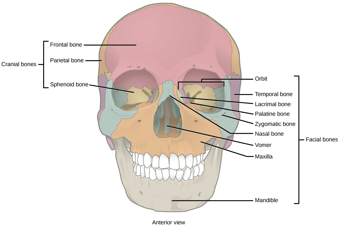
Bones That Form The Orbit
Bones That Form The Orbit - Optic foramen orbital margin (rim): It is our job as ophthalmologists to be able to readily identify these bones and know pretty much every bump, notch, hole, and contour of these bones and what structures pass through, travel along, and attach to these bones. Web there are 7 bones that comprise the orbit. The orbit is made up of portions of both the cranial and the facial skeletal systems. Learn vocabulary, terms, and more with flashcards, games, and other study tools. Web there are seven bones that contribute to the bony orbit: Web the orbit, which protects, supports, and maximizes the function of the eye, is shaped like a quadrilateral pyramid, with its base in plane with the orbital rim. Though small, the orbital bones are quite strong to protect the eye inside the head. The facial bones include 14 bones, with six paired bones and two unpaired bones. Web bones of the orbit and some of the major landmarks.
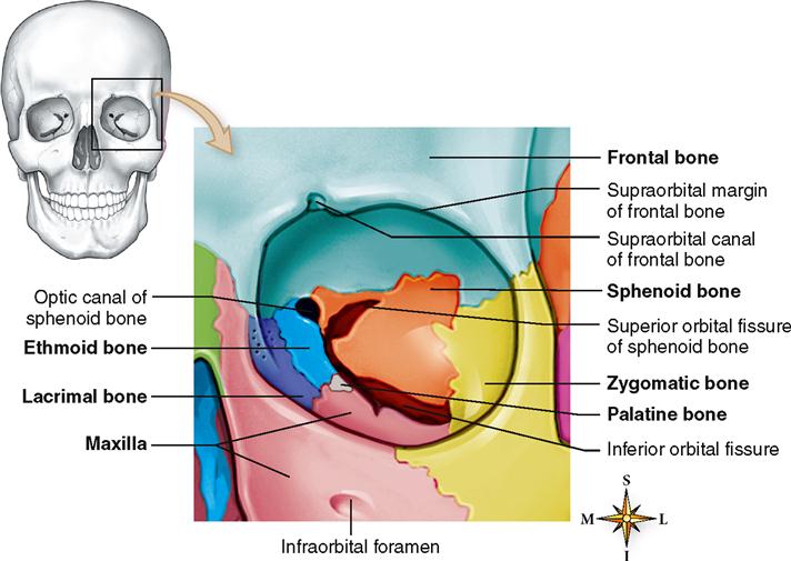
The orbital roof is formed by the lesser wing of the sphenoid bone and the frontal bone. Web portions of six bones form its pyramidal walls: Web bones of the orbit and some of the major landmarks. Web the face is attached anteriorly and consists of two unpaired bones, the vomer and mandibular bones, and six paired bones, the nasal, maxillary, zygomatic, palatine, lacrimal, and inferior turbinate bones. Web the orbit is the bony cavity in the skull that houses the globe of the eye (eyeball), the muscles that move the eye (the extraocular muscles), the lacrimal gland, and the blood vessels and nerves required to supply these structures. Web define bones of the orbit. Web the seven bones that form the orbit: It is our job as ophthalmologists to be able to readily identify these bones and know pretty much every bump, notch, hole, and contour of these bones and what structures pass through, travel along, and attach to these bones. The lateral wall comprises the greater wing of the sphenoid bone and zygomatic bone. The sphenoid and ethmoid bones form mostly via endochondral ossification while the frontal bone is formed by intramembranous ossification.
Web start studying bones that form the orbit part 1. Though small, the orbital bones are quite strong to protect the eye inside the head. The orbit is made up of portions of both the cranial and the facial skeletal systems. Web the face is attached anteriorly and consists of two unpaired bones, the vomer and mandibular bones, and six paired bones, the nasal, maxillary, zygomatic, palatine, lacrimal, and inferior turbinate bones. Web define bones of the orbit. Each of these plays a role in keeping the eyeball protected. Web the facial bones of the skull form the upper and lower jaws, the nose, nasal cavity and nasal septum, and the orbit. Frontal, ethmoid, palatine, lacrimal, maxilla, zygomatic, and sphenoid. Web there are seven bones that contribute to the bony orbit: Web the bony orbits (or eye sockets) are bilateral and symmetrical cavities in the head.

Bones of the orbit Human anatomy and physiology, Anatomy, Orbit anatomy
Seven bones conjoin to form the. Web the following seven bones form the orbit: Formed by the lesser wing of the sphenoid and the frontal bone. What is the function of the orbit? The orbit is made up of portions of both the cranial and the facial skeletal systems.

Bones That Form The Orbit / Orbital Bones Ophthalmology Review
Web there are seven orbital bones that make up this structure: Web start studying bones that form the orbit part 1. Web key facts about bones of the orbit. It is our job as ophthalmologists to be able to readily identify these bones and know pretty much every bump, notch, hole, and contour of these bones and what structures pass.

Bones of orbit lateral wall Human anatomy and physiology, Human
There are 7 bones that form the orbit: The lateral wall comprises the greater wing of the sphenoid bone and zygomatic bone. Web there are seven orbital bones that make up this structure: The orbital roof is formed by the lesser wing of the sphenoid bone and the frontal bone. Web seven bones form each orbit:

The Bony Orbit Borders Contents Fractures TeachMeAnatomy
Each of these plays a role in keeping the eyeball protected. Frontal, ethmoid, palatine, lacrimal, maxilla, zygomatic, and sphenoid. Yellow = frontal bone green = lacrimal bone brown = ethmoid bone blue = zygomatic bone purple = maxillary bone aqua = palatine bone red = sphenoid bone teal = nasal bone (illustrated but not part of the orbit) Orbital plate.

Anatomy bones, Orbit anatomy, Anatomy
Web seven bones form each orbit: Orbital plate of the frontal bone. Lesser wing of the sphenoid bone. Frontal, sphenoid, maxillary, zygomatic, palatine, ethmoid, and lacrimal. Web bones of the orbit and some of the major landmarks.
19.1 Types of Skeletal Systems Concepts of Biology 1st Canadian Edition
Frontal, ethmoid, palatine, lacrimal, maxilla, zygomatic, and sphenoid. The paired bones are the maxilla, palatine, zygomatic, nasal, lacrimal, and inferior nasal conchae bones. Web the bones of the orbit develop via both endochondral and intramembranous ossification. Web there are seven orbital bones that make up this structure: Web the orbit, which protects, supports, and maximizes the function of the eye,.

bones that form the orbit Diagram Quizlet
Web the seven bones that form the orbit: Seven bones conjoin to form the. Formed by the lesser wing of the sphenoid and the frontal bone. In this article, we shall look at the borders, contents and clinical correlations of. Frontal, ethmoid, palatine, lacrimal, maxilla, zygomatic, and sphenoid.

20 best Ophtho images on Pinterest Anatomy, Anatomy reference and
The orbit is a pear shape, with the optic nerve at the stem, and holds approximately 30 cc volume. The facial bones include 14 bones, with six paired bones and two unpaired bones. Learn vocabulary, terms, and more with flashcards, games, and other study tools. Yellow = frontal bone green = lacrimal bone brown = ethmoid bone blue = zygomatic.
Skeletal System Basicmedical Key
Formed by the lesser wing of the sphenoid and the frontal bone. Web seven bones form each orbit: Pars orbitalis of the frontal bone lacrimal bone lamina papyracea of the ethmoid bone orbital process of the zygomatic bone orbital surface of the maxillary bone orbital process of the palatine bone greater and lesser wings and body of the sphenoid bone.

Orbital Bone Anatomy Human Anatomy Diagram Medical anatomy, Human
Bones of the orbit synonyms, bones of the orbit pronunciation, bones of the orbit translation, english dictionary definition of bones of the orbit. The orbit is made up of portions of both the cranial and the facial skeletal systems. Frontal, ethmoid, palatine, lacrimal, maxilla, zygomatic, and sphenoid. Seven bones conjoin to form the. Web let's look at how these seven.
Yellow = Frontal Bone Green = Lacrimal Bone Brown = Ethmoid Bone Blue = Zygomatic Bone Purple = Maxillary Bone Aqua = Palatine Bone Red = Sphenoid Bone Teal = Nasal Bone (Illustrated But Not Part Of The Orbit)
Bones of the orbit synonyms, bones of the orbit pronunciation, bones of the orbit translation, english dictionary definition of bones of the orbit. Web the orbit is the bony cavity in the skull that houses the globe of the eye (eyeball), the muscles that move the eye (the extraocular muscles), the lacrimal gland, and the blood vessels and nerves required to supply these structures. Frontal, ethmoid, palatine, lacrimal, maxilla, zygomatic, and sphenoid. Web the face is attached anteriorly and consists of two unpaired bones, the vomer and mandibular bones, and six paired bones, the nasal, maxillary, zygomatic, palatine, lacrimal, and inferior turbinate bones.
It Is Our Job As Ophthalmologists To Be Able To Readily Identify These Bones And Know Pretty Much Every Bump, Notch, Hole, And Contour Of These Bones And What Structures Pass Through, Travel Along, And Attach To These Bones.
The orbit is made up of portions of both the cranial and the facial skeletal systems. Bones, muscles, arteries, veins and nerves. Seven bones conjoin to form the. In this article, we shall look at the borders, contents and clinical correlations of.
Pars Orbitalis Of The Frontal Bone Lacrimal Bone Lamina Papyracea Of The Ethmoid Bone Orbital Process Of The Zygomatic Bone Orbital Surface Of The Maxillary Bone Orbital Process Of The Palatine Bone Greater And Lesser Wings And Body Of The Sphenoid Bone
Maxilla, frontal bone, zygomatic bone, ethmoid bone, lacrimal bone, sphenoid bone, and palatine bone. The entrance to the globe anteriorly is approximately 35 mm high and 45 mm wide. Web bones of the orbit and some of the major landmarks. They enclose the eyeball and its associated structures.
Web There Are Seven Bones That Contribute To The Bony Orbit:
The lateral wall comprises the greater wing of the sphenoid bone and zygomatic bone. Web names of the bones of the orbit with basic anatomy 7 of the cranial and facial bones contribute to the formation of the orbital cavities, with 3 being cranial bones and the other 4 being facial bones: The orbit is a pear shape, with the optic nerve at the stem, and holds approximately 30 cc volume. The orbital roof is formed by the lesser wing of the sphenoid bone and the frontal bone.